Open Seminar: AI for Science Meetup #2
2026-02-17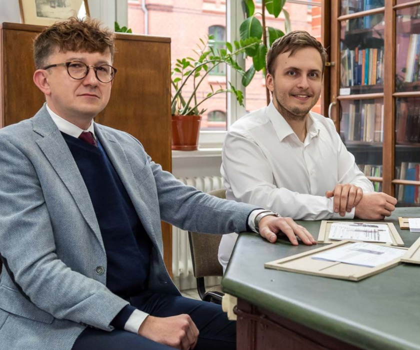
Every scientific journey begins with a question—and with a meeting. Sometimes a single conversation is enough for an idea to gain direction and for doubts to turn into a concrete action plan. In this series, we show how scientific mentoring truly shapes the development of young researchers and how projects combining artificial intelligence in medicine with clinical practice are born.
This was the case with the collaboration between Prof. Piotr Donizy and Bartosz Poniewierka—a medical student who decided to apply AI in histopathological diagnostics.
From Conversation to Research Project: AI and Histopathological Slide Analysis
At the end of last year, Prof. Piotr Donizy was teaching substitute pathology classes for fourth-year medical students. After one session, a student approached him with an intriguing proposal: could artificial intelligence be used to analyze histopathological slides? Could a laser beam detect cancer cells within a fraction of a second?
That conversation initiated their collaboration within the “Young Science” mentoring program.
Bartek didn’t just have an idea—he already had advanced research and well-developed reflections,” admits Prof. Piotr Donizy. “All he needed was support in turning it into a concrete plan, giving it direction, and navigating administrative procedures.
Bartosz Poniewierka combines medicine with computer science. He works in a biotechnology company implementing artificial intelligence solutions while simultaneously developing research projects within the Student Scientific Club Medical Image Analysis and Research in Pathology (MIARP).
At that moment, we needed support in writing grant applications, which we prepared as the Student Scientific Club Medical Image Analysis and Research in Pathology,” says Bartosz Poniewierka. “However, it wasn’t only a technical issue—although that support is invaluable, since we lack experience in navigating bureaucratic barriers. From my perspective, the professor’s experience and extensive knowledge are priceless. Thanks to them, some ideas can grow, and others can be created from scratch.
Beyond the Standard: Mentoring in Science and Research Skill Development
The collaboration between mentor and student goes far beyond formal consultations. Regular discussions—often late in the evening—cover both pathology and advanced technologies, including lasers, data analysis, and the automation of diagnostics.
Prof. Piotr Donizy emphasizes that the relationship works both ways—the mentee’s IT expertise and fresh perspective are equally inspiring. It is a model of academic mentoring that supports intergenerational knowledge transfer.
Their collaboration has resulted in three grant applications:
-
a microgrant in microbiology (using Raman spectroscopy for pathogen identification),
-
a project within the Student Activity Fund (FAST) for retinal screening,
-
an application to the “Student Scientific Clubs Create Innovations” program.
The project “InsightEye: Look into the Eyes” received funding within FAST, confirming the innovation potential of student research teams.
Raman Spectroscopy and AI in Skin Cancer Diagnostics
The most ambitious project focuses on creating a system that combines Raman spectroscopy with automated computer analysis in the diagnosis of skin cancers.
This project could introduce in Poland a solution comparable to leading global centers: a system combining highly advanced biopsy sample examination with automated computer analysis,” emphasizes Bartosz Poniewierka. “It is an idea that has the potential to fundamentally change how we look at a specimen—moving from a microscope image to data that can be objectively compared and interpreted. The goal is to support the diagnosis of malignant skin cancers by creating a database and then developing artificial intelligence software capable of evaluating slide scans and indicating whether cancer cells are present and what type of lesion it is.
The project includes, among others:
-
melanoma,
-
basal cell carcinoma,
-
squamous cell carcinoma,
-
Merkel cell carcinoma.
The team plans to conduct large-scale Raman spectral measurements, build a structured database, and train AI algorithms that remain robust even for rare variants of lesions.
Raman spectroscopy enables tissue composition analysis using laser light. Each tissue generates a characteristic “fingerprint”—a spectrum that differs depending on the presence of disease. Combining this technology with artificial intelligence algorithms may enhance the objectivity and reproducibility of diagnostics.
The students have already developed procedures to remove autofluorescence and to technologically prepare substrates that minimize measurement interference. The results of their research will be published soon.
This is precisely why the students’ idea carries such clinical significance: it is not just about creating another program, but about building competencies and infrastructure that allow these analyses to be performed locally—without the costly and time-consuming ‘export’ of diagnostics and research abroad,” says Prof. Piotr Donizy. “What is innovative here is not only the use of AI, but above all the attempt to combine the Raman ‘fingerprint’ of tissue with classical pathology in a way that can deliver repeatable, measurable, and clinically useful results. Introducing such a solution would mean a real opportunity for faster and more objective support of diagnostic decisions and, consequently, shortening the patient’s path from suspected disease to the initiation of treatment, particularly in rare conditions such as amyloidoses. Equally important, it would represent a unique nationwide example of integrating Raman slide scanning with AI-based analysis, developed bottom-up by a student team but with ambitions to scale and implement it more broadly.
SERS, Antibiotic Resistance and the Future of Microbiological Diagnostics
The applications of Raman spectroscopy extend beyond pathology.
However, Raman spectroscopy is not limited to pathology alone,” emphasizes Bartosz Poniewierka. “Together with dr hab. Emil Albert Paluch, prof. UMW, we are working on developing a special SERS (Surface Enhanced Raman Spectroscopy) substrate that will enhance the signal from bacteria under laser illumination. Based on the results obtained in this way, we want to create a database that will initially allow species-level pathogen differentiation and, in the longer term, provide information about antimicrobial resistance spectra—without the need for time-consuming culture. Considering the growing problem of antibiotic resistance, this system could accelerate the implementation of targeted therapy. These are very long-term plans, with a perspective of more than a decade, but we believe we will achieve them.
Pathology as a Space for New Technologies
Although Bartosz Poniewierka initially planned to pursue a surgical specialty, today he increasingly sees his future in pathology—a field that is becoming one of the key areas for the development of modern data-driven medical diagnostics.
It is a story about how AI in medicine, Raman spectroscopy, and the engagement of a student scientific club can meet the experience of a mentor—and together create a project with the potential to bring real change to Polish oncological diagnostics.


